Beranda
/ Anatomy Of Chest / Human Anatomy Illustration Chest Rib Cage Vascular System 3d Illustration Stock Photo Alamy : The tissue adjacent to the aorta is the.
Anatomy Of Chest / Human Anatomy Illustration Chest Rib Cage Vascular System 3d Illustration Stock Photo Alamy : The tissue adjacent to the aorta is the.
Insurance Gas/Electricity Loans Mortgage Attorney Lawyer Donate Conference Call Degree Credit Treatment Software Classes Recovery Trading Rehab Hosting Transfer Cord Blood Claim compensation mesothelioma mesothelioma attorney Houston car accident lawyer moreno valley can you sue a doctor for wrong diagnosis doctorate in security top online doctoral programs in business educational leadership doctoral programs online car accident doctor atlanta car accident doctor atlanta accident attorney rancho Cucamonga truck accident attorney san Antonio ONLINE BUSINESS DEGREE PROGRAMS ACCREDITED online accredited psychology degree masters degree in human resources online public administration masters degree online bitcoin merchant account bitcoin merchant services compare car insurance auto insurance troy mi seo explanation digital marketing degree floridaseo company fitness showrooms stamfordct how to work more efficiently seowordpress tips meaning of seo what is an seo what does an seo do what seo stands for best seotips google seo advice seo steps, The secure cloud-based platform for smart service delivery. Safelink is used by legal, professional and financial services to protect sensitive information, accelerate business processes and increase productivity. Use Safelink to collaborate securely with clients, colleagues and external parties. Safelink has a menu of workspace types with advanced features for dispute resolution, running deals and customised client portal creation. All data is encrypted (at rest and in transit and you retain your own encryption keys. Our titan security framework ensures your data is secure and you even have the option to choose your own data location from Channel Islands, London (UK), Dublin (EU), Australia.
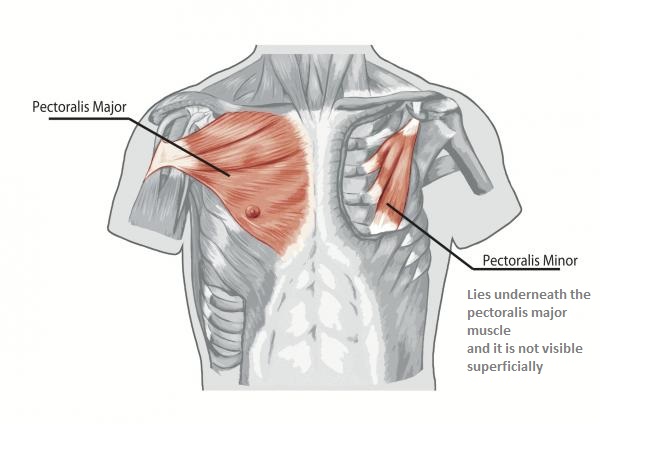
Anatomy Of Chest / Human Anatomy Illustration Chest Rib Cage Vascular System 3d Illustration Stock Photo Alamy : The tissue adjacent to the aorta is the.. Structures of the heart such as the right ventricle (3), intraventricular septum (4), left ventricular free wall (5) and papillary muscles (arrow) are clearly seen. It provides access to ct images in the axial plane, allowing the user to learn and review the lung anatomy interactively. As with all parts of the body, the anatomy and physiology of the chest wall are intimately intertwined. The epidermis is the outermost layer that provides a protective, waterproof seal over the body. The major muscle in the chest is the pectoralis major.
However, the classical anatomical descriptions in textbooks make it difficult to gain full mastery of this subject, because the books usually deal with its elements separately. A good radiologist knows the anatomy because knowing where structures normally live and recognizing the location of an abnormality helps to make or narrow the differential diagnosis. Radiology basics of chest ct anatomy with annotated coronal images and scrollable axial images to help medical students and junior doctors learning anatomy. The major muscle in the chest is the pectoralis major. Your sternum protects the organs of your torso from injury and also serves as a.
Pertinent Surgical Anatomy Of The Thorax And Mediastinum Clinical Gate from clinicalgate.com This atlas is a comprehensive and affordable learning tool for medical students and residents and especially for radiologists and pneumologists. This chapter is an abbreviated review of thoracic anatomy as seen on chest radiographs and computed tomography (ct) of the chest. The pec major) is the one that commands the most real estate. It provides access to ct images in the axial plane, allowing the user to learn and review the lung anatomy interactively. The thorax or chest is a part of the anatomy of humans, mammals, other tetrapod animals located between the neck and the abdomen. Anatomy of right side chest pain. An overview of the anatomy visible in a transverse computed axial tomographical image of the thorax (and part of the abdomen) performed with intravenous cont. However, the classical anatomical descriptions in textbooks make it difficult to gain full mastery of this subject, because the books usually deal with its elements separately.
The human thorax includes the thoracic cavity and the thoracic wall.
It provides protection to vital organs (eg, heart and major vessels, lungs, liver) and provides stability for movement. Your sternum protects the organs of your torso from injury and also serves as a. 12 cm (5 in) in length, 8 cm (3.5 in) wide, and 6 cm (2.5 in) in thickness. Skandalakis chest wall embryogenesis the muscles of the chest develop from the somites found in the mesoderm. Intravenous contrast is seen in the left ventricle (1) and descending aorta (2). Swensen fund for innovation in teaching. Radiology basics of chest ct anatomy with annotated coronal images and scrollable axial images to help medical students and junior doctors learning anatomy. Related posts of anatomy of the chest area anatomy of the ankle and foot. Here, we break down the anatomy of your chest muscles. Thoracic cavity, also called chest cavity, the second largest hollow space of the body. In insects, crustaceans, and the extinct trilobites, the thorax is one of the three main divisions of the creature's body, each of which is in turn composed of multiple segments. The chest is the area of origin for many of the body's systems as it houses organs such as the heart, esophagus, trachea, lungs, and thoracic diaphragm. The pec major) is the one that commands the most real estate.
In insects, crustaceans, and the extinct trilobites, the thorax is one of the three main divisions of the creature's body, each of which is in turn composed of multiple segments. About the 6th week, the somites differentiate into the sclerotomes and the dermatomyotomes. A good radiologist knows the anatomy because knowing where structures normally live and recognizing the location of an abnormality helps to make or narrow the differential diagnosis. The epidermis is the outermost layer that provides a protective, waterproof seal over the body. As with all parts of the body, the anatomy and physiology of the chest wall are intimately intertwined.
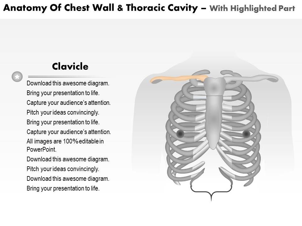
Diagram Tattoo Chest Diagram Full Version Hd Quality Chest Diagram Carbeltdiagrams Seewhatimean It from bodybuilding-wizard.com It's also sometimes referred to as the breastbone. It is enclosed by the ribs, the vertebral column, and the sternum, or breastbone, and is separated from the abdominal cavity (the body's largest hollow space) by a muscular and membranous partition, the diaphragm. Computed tomography (ct) of the chest can detect pathology that may not show up on a conventional chest radiograph(1). Here, we break down the anatomy of your chest muscles. Intravenous contrast is seen in the left ventricle (1) and descending aorta (2). This chapter is an abbreviated review of thoracic anatomy as seen on chest radiographs and computed tomography (ct) of the chest. How to view the anatomical labels. The circulatory system does most of its work.
A typical heart is approximately the size of your fist:
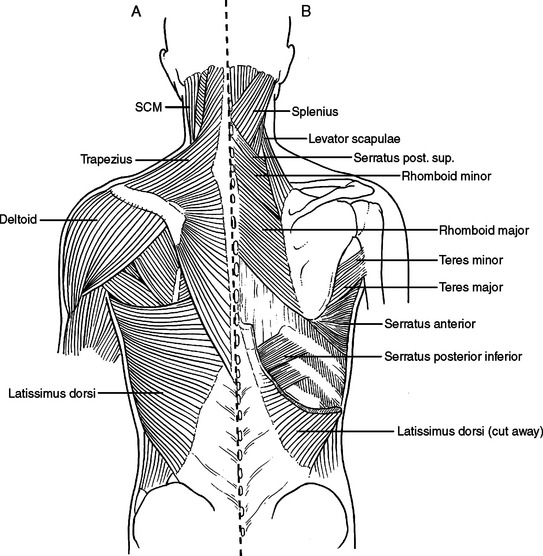
The chest wall is comprised of skin, fat, muscles, and the thoracic skeleton. 12 cm (5 in) in length, 8 cm (3.5 in) wide, and 6 cm (2.5 in) in thickness. Your sternum protects the organs of your torso from injury and also serves as a. As with all parts of the body, the anatomy and physiology of the chest wall are intimately intertwined. Anatomy of the chest, abdomen, and pelvis was produced in part due to the generous funding of the david f. The human thorax includes the thoracic cavity and the thoracic wall. Browse 2,531 female chest anatomy stock photos and images available, or start a new search to explore more stock photos and images. Structures of the heart such as the right ventricle (3), intraventricular septum (4), left ventricular free wall (5) and papillary muscles (arrow) are clearly seen. Radiology basics of chest ct anatomy with annotated coronal images and scrollable axial images to help medical students and junior doctors learning anatomy. This article focuses on the unique structural characteristics in … Applied anatomy of the chest wall and mediastinum petros mirilas michael e. How to view the anatomical labels. Of the two chest muscles, the pectoralis major (a.k.a.
This atlas is a comprehensive and affordable learning tool for medical students and residents and especially for radiologists and pneumologists. To carry out the unique functions performed by the chest wall, the anatomic structures are formed precisely for maximal efficiency. The major muscle in the chest is the pectoralis major. Related posts of anatomy of the chest area anatomy of the ankle and foot. The epidermis is the outermost layer that provides a protective, waterproof seal over the body.
0514 Anatomy Of Chest Wall And Thoracic Cavity Medical Images For Powerpoint Graphics Presentation Background For Powerpoint Ppt Designs Slide Designs from www.slideteam.net To carry out the unique functions performed by the chest wall, the anatomic structures are formed precisely for maximal efficiency. Related posts of anatomy of the chest area anatomy of the ankle and foot. Computed tomography (ct) of the chest can detect pathology that may not show up on a conventional chest radiograph(1). Applied anatomy of the chest wall and mediastinum petros mirilas michael e. The circulatory system does most of its work. The chest wall is comprised of skin, fat, muscles, and the thoracic skeleton. 12 cm (5 in) in length, 8 cm (3.5 in) wide, and 6 cm (2.5 in) in thickness. The pec major) is the one that commands the most real estate.
Here, we break down the anatomy of your chest muscles.
To carry out the unique functions performed by the chest wall, the anatomic structures are formed precisely for maximal efficiency. Browse 2,531 female chest anatomy stock photos and images available, or start a new search to explore more stock photos and images. Your sternum protects the organs of your torso from injury and also serves as a. Chest a man's chest — like the rest of his body — is covered with skin that has two layers. A typical heart is approximately the size of your fist: The chest is the area of origin for many of the body's systems as it houses organs such as the heart, esophagus, trachea, lungs, and thoracic diaphragm. An overview of the anatomy visible in a transverse computed axial tomographical image of the thorax (and part of the abdomen) performed with intravenous cont. As with all parts of the body, the anatomy and physiology of the chest wall are intimately intertwined. It's also sometimes referred to as the breastbone. The chest anatomy includes the pectoralis major, pectoralis minor and the serratus anterior. Anatomy of right side chest pain. Of the two chest muscles, the pectoralis major (a.k.a. About the 6th week, the somites differentiate into the sclerotomes and the dermatomyotomes.